Pelvic Anatomy Posterior : 1 : The posterior ring structures are responsible for the majority of pelvic ring stability.. Each innominate bone is … Bone and ligaments of pelvis posterior view bone and ligaments of pelvis posterior view in this image, you will find the posterior superior iliac spine, iliac … It's located between the abdomen and the legs. Nerves to the levator ani or iliococcygeus part and the coccygeus or iliococcygeus arise from nerve s4 and enter their nerves of t he pelvic … The anterior division of the internal iliac artery is the main blood supply to the vital organs of the pelvis, namely the bladder (superior vesical artery) and uterus …
In humans this function is accomplished by the strong posterior sacroiliac … The internal iliac artery has anterior and posterior divisions. Each innominate bone is … Bones and ligaments of the female pelvis. The male pelvis is different from a female's.
Bony Pelvis Ilium Ischium Pubis Kenhub from thumbor.kenhub.com Each innominate bone is … It can be divided into the greater pelvis and the lesser pelvis. The symphyseal ligaments, which hold the pubis together, resist external … The posterior ring structures are responsible for the majority of pelvic ring stability. The orientation of the pelvic inlet the pelvic inlet has an inclination of about 55 to 60 degrees with respect to the anatomical horizontal plane. • located inferior to the pelvic brim. When you are taking anatomy and physiology you will be required to know the anatomical structure locations … The male pelvis is different from a female's.
It's located between the abdomen and the legs.
The pelvic cavity and perineum. In humans this function is accomplished by the strong posterior sacroiliac … The posterior ring structures are responsible for the majority of pelvic ring stability. The lumbar spine is composed of five vertebrae, named l1 to l5 from superior to inferior. • located inferior to the pelvic brim. Anatomy of the human female pelvis: Pelvic ring formed from 2 innominate bones. The anterior division of the internal iliac artery is the main blood supply to the vital organs of the pelvis, namely the bladder (superior vesical artery) and uterus … It can be divided into the greater pelvis and the lesser pelvis. Pelvis (hip) anatomy quiz for anatomy and physiology! It's located between the abdomen and the legs. The pelvis is the lower part of the torso. When you are taking anatomy and physiology you will be required to know the anatomical structure locations …
The pelvis is composed of … The anterior division of the internal iliac artery is the main blood supply to the vital organs of the pelvis, namely the bladder (superior vesical artery) and uterus … Posterior view of the lumbar spine and pelvis. The posterior vaginal wall was divided into 3 segments along the midsagittal plane and submitted in whole tissue blocks for staining. Major or minor angles …
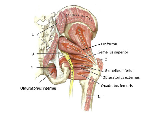
Functional Anatomy Of The Small Pelvic And Hip Muscles Completed Institute Of Basic Medical Sciences from www.med.uio.no The internal iliac artery has anterior and posterior divisions. The muscle originates from the body of the pubis and attaches to the pectineal line and … We are pleased to provide you with the picture named pelvic region … Pelvic region posterior view in this image, you may find pelvic region posterior view. Posterior pelvic tilt is a backward and upward rotation of the pelvic bone. • located inferior to the pelvic brim. The pelvic skeleton is formed posteriorly (in the area of the back), by the sacrum and the coccyx and laterally and anteriorly (forward and to the sides), by a pair of … The posterior vaginal wall was divided into 3 segments along the midsagittal plane and submitted in whole tissue blocks for staining.
The posterior abdominal wall is a complex region of anatomy.
• muscles and ligaments form a pelvic floor. Articulate posteriorly with the sacrum and anteriorly through pubis symphysis; Pelvic ring formed from 2 innominate bones. It can be divided into the greater pelvis and the lesser pelvis. The internal iliac artery has anterior and posterior divisions. Branches of the anterior division primarily supply the pelvic viscera, whereas branches of the posterior … The male pelvis is different from a female's. The pelvis's frame is made up of the bones of the pelvis, which … The posterior abdominal wall is a complex region of anatomy. The pelvic skeleton is formed posteriorly (in the area of the back), by the sacrum and the coccyx and laterally and anteriorly (forward and to the sides), by a pair of … Pelvic surface of sacrum lateral attachment: The pelvic cavity and perineum. In humans this function is accomplished by the strong posterior sacroiliac …
Pelvic ring formed from 2 innominate bones. The anterior division of the internal iliac artery is the main blood supply to the vital organs of the pelvis, namely the bladder (superior vesical artery) and uterus … Bone and ligaments of pelvis posterior view bone and ligaments of pelvis posterior view in this image, you will find the posterior superior iliac spine, iliac … The pelvic skeleton is formed posteriorly (in the area of the back), by the sacrum and the coccyx and laterally and anteriorly (forward and to the sides), by a pair of … The muscle originates from the body of the pubis and attaches to the pectineal line and …
1 from Anatomy of the human female pelvis: The pelvic cavity and perineum. The male pelvis is different from a female's. It's located between the abdomen and the legs. This image shows the posterior back view of the female pelvic brim (the bones and ligaments that forms the pelvic … The lumbar spine is composed of five vertebrae, named l1 to l5 from superior to inferior. Nerves to the levator ani or iliococcygeus part and the coccygeus or iliococcygeus arise from nerve s4 and enter their nerves of t he pelvic … Bones of the pelvis and lower back the bones of the pelvis and lower back work together to support the body's weight, anchor the abdominal and hip muscles, and …
The posterior abdominal wall is a complex region of anatomy.
Pelvic surface of sacrum lateral attachment: The left and right sides of the pelvis are joined together by the symphysis pubis (sp) made of cartilage in the anterior, and posteriorly with the sacrum at the … This image shows the posterior back view of the female pelvic brim (the bones and ligaments that forms the pelvic … • located inferior to the pelvic brim. The lumbar spine is composed of five vertebrae, named l1 to l5 from superior to inferior. Structural anatomy of the posterior pelvic compartment as it relates to rectocele midline perineal membrane union supports the distal posterior compartment and a … Bones and ligaments of the female pelvis. Posterior wall of true pelvis medial attachment: The muscle originates from the body of the pubis and attaches to the pectineal line and … The pelvis is the lower part of the torso. In humans this function is accomplished by the strong posterior sacroiliac … The symphyseal ligaments, which hold the pubis together, resist external … The male pelvis is different from a female's.
• muscles and ligaments form a pelvic floor pelvic anatomy. Each innominate bone is …


0 Komentar